
The Radiology Ordering Guide provides evidence-based recommendations for selecting appropriate imaging exams, ensuring optimal patient care and diagnostic accuracy while minimizing unnecessary procedures․
1․1 Purpose and Scope
The Radiology Ordering Guide is designed to assist healthcare providers in selecting the most appropriate imaging procedures․ Its purpose is to optimize diagnostic accuracy, minimize unnecessary exams, and enhance patient outcomes․ The scope includes evidence-based recommendations for various imaging modalities, patient preparation, contrast agents, and special considerations like pediatric and geriatric imaging․ It serves as a comprehensive resource for both common and complex clinical scenarios, ensuring high-quality care․
1․2 Importance of Appropriate Imaging
Appropriate imaging is crucial for accurate diagnosis, effective treatment planning, and minimizing healthcare costs․ It reduces unnecessary radiation exposure and avoids repeat procedures․ By adhering to evidence-based guidelines, clinicians ensure patient safety and optimal outcomes․ Proper imaging also helps in avoiding complications and improves overall quality of care․ It aligns with best practices, supporting informed decision-making and efficient resource utilization․
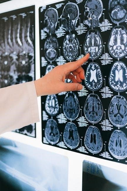
Understanding Radiology Basics
Understanding radiology basics involves familiarity with common radiographic projections, positioning techniques, and essential imaging modalities like X-ray, CT, MRI, and Ultrasound․ These fundamentals are critical for accurate diagnoses and effective treatment planning․
2․1 Common Radiographic Projections and Positioning
Common radiographic projections include anteroposterior (AP) and lateral views, ensuring accurate visualization of anatomical structures․ Proper patient positioning is critical to obtain high-quality images, minimizing distortions and ensuring diagnostic accuracy; Standardized techniques, such as supine or upright positions, are used based on the body region being imaged․ Clear communication between technologists and patients ensures correct alignment, optimizing image quality and reducing the need for repeat exams․ Proper positioning enhances diagnostic confidence and patient safety․
2․2 Essential Imaging Modalities (X-ray, CT, MRI, Ultrasound)
X-ray is the most common modality, ideal for bone and lung imaging due to its accessibility and low cost․ CT scans provide detailed cross-sectional images, especially for trauma and abdominal pathologies․ MRI excels in soft tissue visualization, such as brain and joint disorders, while ultrasound offers real-time imaging without radiation, perfect for obstetrics and vascular assessments․ Each modality has unique strengths, and selecting the right one depends on the clinical scenario, patient condition, and diagnostic needs, guided by evidence-based protocols like the ACR Appropriateness Criteria․

Clinical Indications for Radiology Exams
This section provides evidence-based guidelines for selecting the most appropriate imaging exams for common medical conditions, trauma, and specialized areas like women’s imaging, ensuring optimal patient outcomes and minimizing unnecessary procedures․
3․1 Guidelines for Common Medical Conditions
This section outlines evidence-based criteria for selecting imaging exams based on common medical conditions, ensuring appropriate use of radiology․ For conditions like chest pain, respiratory issues, or abdominal discomfort, specific imaging modalities such as X-ray, CT, or ultrasound are recommended․ It also covers chronic conditions, including cardiovascular and musculoskeletal diseases, providing guidance on when MRI or PET/CT is most effective․ The goal is to balance diagnostic accuracy with patient safety and cost-effectiveness, optimizing care through tailored imaging strategies․
3․2 Trauma Imaging Best Practices
Trauma imaging requires rapid, accurate assessment to guide emergency care․ Guidelines recommend prioritizing CT for blunt trauma evaluation, especially for head, chest, and abdominal injuries․ Avoid unnecessary delays and ensure patient stability before imaging․ For penetrating trauma, ultrasound is often the first-line tool․ Always consider radiation exposure risks, particularly in pediatric patients․ Adhere to ACR Appropriateness Criteria for trauma-specific protocols to optimize diagnostic yield and patient outcomes, ensuring timely and effective management of critically injured patients․
3․3 Women’s Imaging (Breast, Pelvic, and Obstetric Imaging)
Women’s imaging focuses on breast, pelvic, and obstetric conditions․ Guidelines recommend mammography and ultrasound for breast screening and diagnostic evaluation․ Pelvic MRI is optimal for assessing indeterminate adnexal masses․ Obstetric ultrasound is crucial for fetal evaluation and placental assessment․ Contrast-enhanced MRI is preferred for complex pelvic pathology․ Ensure adherence to ACR Appropriateness Criteria for tailored imaging approaches, minimizing radiation and contrast use, especially during pregnancy and breastfeeding, to optimize diagnostic accuracy and patient safety․

Contrast Agents in Radiology
Contrast agents enhance image quality, aiding in diagnosis․ Common types include iodine-based for CT and gadolinium-based for MRI․ Use requires precautions due to contraindications and allergies․
4․1 Types of Contrast Agents (Iodine-Based, Gadolinium-Based)
Contrast agents are substances used to enhance image clarity․ Iodine-based agents are commonly used in CT scans and X-rays, while gadolinium-based agents are typically used in MRI․ Both types help highlight specific tissues or structures, improving diagnostic accuracy․ However, precautions are necessary, as iodine-based agents can cause allergic reactions, and gadolinium-based agents have rare but serious side effects․ Selection depends on patient history, medical conditions, and the desired diagnostic outcome․
4․2 Contraindications and Precautions
Contraindications for contrast agents vary by type․ Iodine-based contrasts are contraindicated in patients with severe allergies or prior anaphylactic reactions․ Gadolinium-based agents are avoided in those with severe kidney disease due to the risk of nephrogenic systemic fibrosis․ Pregnant or breastfeeding women require careful consideration․ Precautions include assessing renal function, screening for allergies, and monitoring for adverse reactions․ Radiologists play a key role in evaluating individual risks and ensuring safe contrast administration․
4․3 Best Practices for Contrast Administration
Best practices for contrast administration include assessing patient history for allergies and renal function․ Ensure informed consent and pre-procedure hydration, especially for high-risk patients․ Use test doses for iodine-based contrasts when history is unclear․ Monitor patients during and after administration for adverse reactions․ Document all procedures and ensure adherence to standardized protocols․ Radiologists should tailor contrast use to individual needs, balancing diagnostic benefits with potential risks․ Proper training and emergency preparedness are essential for safe contrast administration․

Patient Preparation for Radiology Exams
Proper preparation ensures accurate results, patient safety, and effective imaging outcomes․ Guidelines vary by modality, covering fasting, medications, and other specific requirements to optimize diagnostic quality․
5․1 General Preparation Guidelines
General preparation involves fasting for certain exams, avoiding medications that could interfere, and removing metal objects․ Patients should hydrate unless instructed otherwise․ For contrast exams, ensure no allergies․ Provide clear instructions on clothing and arrival times to streamline the process․ Special considerations include pregnancy, breastfeeding, and renal impairment; Adherence to these guidelines ensures safety, optimal image quality, and accurate diagnostic results․ Proper preparation minimizes delays and retakes, improving patient outcomes and operational efficiency․
5․2 Special Considerations (Pregnancy, Breastfeeding, Allergies)
For pregnant patients, avoid exams with ionizing radiation unless essential․ Breastfeeding mothers may need to discontinue feeding temporarily based on institutional guidelines․ Allergies, especially to contrast agents, require careful assessment․ Premedication may be necessary for allergic patients․ Always consider alternative imaging modalities when contraindications exist․ Clear documentation of these considerations ensures patient safety and optimal imaging outcomes․ Adherence to these guidelines minimizes risks and enhances diagnostic accuracy․

Imaging Protocols and Optimization
This section covers standardized imaging protocols, customization for specific patient needs, and strategies for monitoring and adjusting imaging parameters to ensure optimal diagnostic outcomes and patient safety․
6․1 Standardized Imaging Protocols
Standardized imaging protocols ensure consistency and quality in radiology exams by defining optimal parameters for common procedures․ These protocols reduce variability and ensure that imaging meets diagnostic requirements while minimizing radiation exposure․ They are developed based on evidence-based guidelines and professional recommendations, tailored to specific modalities like CT, MRI, and X-ray․ By following these protocols, radiologists and technologists can achieve high-quality images, improving diagnostic accuracy and patient outcomes․ Regular updates ensure alignment with advancements in technology and clinical practices․
6․2 Customizing Protocols for Specific Patient Needs
Customizing imaging protocols for specific patient needs ensures personalized care and optimal diagnostic outcomes․ Factors such as age, condition severity, allergies, and renal impairment guide adjustments․ For example, reducing contrast agents in patients with renal issues or avoiding iodine-based agents in allergic individuals․ Pregnant or breastfeeding patients may require modified protocols to ensure safety․ Referring clinicians and radiologists collaborate to tailor exams, balancing diagnostic accuracy with patient safety; This approach enhances image quality and minimizes risks, ensuring exams are patient-centric and effective․
6․3 Monitoring and Adjusting Imaging Parameters
Monitoring and adjusting imaging parameters during procedures ensures optimal image quality and patient safety․ Real-time adjustments, such as modifying radiation dose or contrast agent timing, are critical․ Technologists and radiologists collaborate to refine settings based on patient anatomy and diagnostic needs․ Continuous feedback loops help identify areas for improvement, ensuring protocols remain effective and patient-centric․ Regular review of imaging outcomes fosters ongoing refinement, enhancing diagnostic accuracy while minimizing risks․

Diagnostic Imaging Procedures
This section details advanced imaging techniques like mammography, MRI for adnexal masses, and nuclear medicine protocols, ensuring accurate diagnosis and effective treatment planning for diverse clinical scenarios․
7․1 Mammography and Breast Ultrasound
Mammography is the primary screening tool for breast cancer, particularly for women over 40, offering early detection of abnormalities․ Breast ultrasound is often used as a complementary tool, especially for younger women with dense breast tissue or to further evaluate suspicious mammography findings․ Both modalities are essential for accurate diagnosis, with mammography providing a broader overview and ultrasound offering detailed tissue assessment․ Their combined use enhances diagnostic accuracy, ensuring timely and appropriate patient care․
7․2 MRI for Indeterminate Adnexal Masses
MRI is a valuable tool for evaluating indeterminate adnexal masses identified on ultrasound, particularly in premenopausal women․ It provides detailed tissue characterization, helping differentiate benign from malignant lesions․ Contrast-enhanced MRI enhances diagnostic accuracy by improving lesion visualization․ The ESUR guidelines recommend an algorithmic approach, utilizing MRI to guide further management․ This modality is especially useful when ultrasound findings are inconclusive, offering a non-invasive means to assess complex adnexal pathology effectively․
7․4 Nuclear Medicine and PET/CT Protocols
Nuclear medicine and PET/CT protocols play a critical role in diagnosing and managing various diseases, offering functional and molecular imaging insights․ Guidelines emphasize optimized imaging times and tracer dosages to ensure diagnostic accuracy․ For instance, 18F-fluciclovine PET/CT is recommended for prostate cancer staging due to its high sensitivity․ Adherence to standardized protocols minimizes radiation exposure and enhances image quality, ensuring safe and effective patient care while avoiding unnecessary procedures․

Special Considerations in Radiology
Special considerations in radiology involve tailored approaches for pediatric, geriatric, and patients with renal impairment, ensuring safe and effective imaging while adhering to specific guidelines and precautions․
8․1 Pediatric Imaging
Pediatric imaging requires careful consideration of radiation exposure, patient cooperation, and appropriate protocol selection․ Using low-dose techniques and age-specific guidelines ensures diagnostic quality while minimizing risks․ Communication with parents and child-friendly environments help reduce anxiety; Specialized training for technologists and radiologists is crucial for optimal outcomes․ Regular updates to protocols based on advancements in technology and evidence-based practices further enhance safety and effectiveness in pediatric radiology․
8․2 Geriatric Imaging
Geriatric imaging focuses on addressing age-related conditions while considering comorbidities and mobility challenges․ Techniques like low-dose CT and MRI are often preferred to minimize risks․ Clear communication and patient-centered care are essential, as elderly patients may have hearing or cognitive impairments; Radiologists should prioritize quick exam times and comfort to reduce stress․ Collaboration with geriatricians ensures tailored imaging approaches, optimizing diagnostic accuracy and patient safety․
8․3 Imaging in Patients with Renal Impairment
Imaging in patients with renal impairment requires careful selection to avoid nephrotoxicity․ Contrast agents like iodine and gadolinium should be used cautiously, with alternatives like ultrasound preferred when possible․ MRI with gadolinium is contraindicated in severe renal failure․ CT scans without contrast are often recommended․ Monitoring renal function before and after imaging is crucial․ Consultation with nephrologists may be necessary for complex cases to ensure safe and effective diagnostic outcomes while minimizing risks․

Clinical Decision Support Systems
Clinical Decision Support Systems (CDSS) guide appropriate imaging selections using evidence-based criteria like ACR Appropriateness Criteria, enhancing diagnostic accuracy and reducing unnecessary exams while improving patient care․
9․1 Role of ACR Appropriateness Criteria
The ACR Appropriateness Criteria (ACR AC) provide evidence-based guidelines for selecting the most suitable imaging procedures for specific clinical conditions․ These criteria are developed by expert panels and updated regularly to reflect current medical knowledge and technology․ By integrating ACR AC into clinical decision support systems, healthcare providers can ensure that imaging orders align with best practices, improve diagnostic accuracy, and reduce unnecessary examinations․ This approach enhances patient outcomes while optimizing resource utilization and reducing healthcare costs․
9․2 Evidence-Based Guidelines
Evidence-based guidelines are systematically developed recommendations that assist referring physicians in selecting the most appropriate imaging procedures․ These guidelines are grounded in scientific evidence and expert consensus, ensuring that imaging decisions are aligned with current best practices․ By integrating evidence-based guidelines into clinical workflows, healthcare providers can enhance diagnostic accuracy, reduce unnecessary procedures, and improve patient outcomes․ Regular updates ensure these guidelines remain relevant and effective in advancing radiology practice․

Legal and Ethical Considerations
Legal and ethical considerations in radiology emphasize informed consent, patient rights, radiation safety, and confidentiality․ Adhering to these principles ensures compliant and respectful patient care․
10․1 Informed Consent and Patient Rights
Informed consent ensures patients are fully aware of the risks, benefits, and alternatives of radiology procedures․ Respecting patient autonomy, dignity, and confidentiality is paramount․ Clinicians must provide clear, understandable information, allowing patients to make informed decisions․ Patient rights include the freedom to refuse or withdraw consent at any time․ Radiology practices must uphold ethical standards, ensuring transparency and adherence to legal requirements, fostering trust between patients and healthcare providers․
10․2 Radiation Safety and Dose Optimization
Radiation safety and dose optimization are critical to minimize risks while maintaining diagnostic quality․ The ALARA principle (As Low As Reasonably Achievable) guides practices to ensure patient exposure is justified and optimized․ Techniques like adjusting exposure factors, using shielding, and employing modern imaging technologies help reduce radiation doses․ Regular monitoring and adherence to evidence-based guidelines, such as the ACR Appropriateness Criteria, further enhance patient safety and overall care quality․
Continuous Improvement in Radiology
Continuous improvement in radiology involves ongoing education, adopting new technologies, and adhering to evidence-based guidelines to enhance diagnostic accuracy and patient care quality․
11․1 Quality Assurance and Quality Control
Quality assurance and quality control in radiology ensure high standards of imaging and diagnostic accuracy․ Regular audits, adherence to guidelines, and feedback mechanisms help identify and correct discrepancies․ These processes reduce repeat procedures, enhance patient safety, and improve overall efficiency․ Continuous monitoring of imaging protocols and equipment performance is essential for maintaining optimal results;
11․2 Feedback Mechanisms for Referring Clinicians
Feedback mechanisms are crucial for improving radiology ordering practices․ Clinicians receive timely, structured reports with radiologist recommendations, ensuring clear communication․ Standardized feedback formats enhance understanding and implementation․ Regular updates on imaging appropriateness and diagnostic accuracy help refine ordering decisions․ This iterative process fosters collaboration, reducing unnecessary exams and optimizing resource use while improving patient outcomes and clinician confidence in radiology services․
The Radiology Ordering Guide ensures appropriate imaging, enhancing diagnostic accuracy and patient care․ It promotes evidence-based practices, fostering collaboration and continuous improvement in radiology services․
12․1 Summary of Key Takeaways
The Radiology Ordering Guide emphasizes evidence-based imaging practices to ensure optimal patient care․ It provides clear guidelines for selecting appropriate exams, balancing diagnostic accuracy with patient safety․ By adhering to these recommendations, clinicians can enhance outcomes while reducing unnecessary procedures․ The guide also highlights the importance of staying updated with advancements in radiology to continuously improve care․ Effective use of imaging resources is crucial for delivering high-quality patient outcomes․
12․2 Future Directions in Radiology Ordering
Future advancements in radiology ordering will focus on integrating AI and machine learning to enhance decision-making․ Cloud-based solutions will optimize image storage and accessibility․ Emphasis will be placed on standardized protocols and patient-centered care․ Continuous updates to guidelines, such as those from the ACR, will ensure evidence-based practices․ Improved data management and radiation dose optimization will remain priorities․ These innovations aim to enhance diagnostic accuracy, streamline workflows, and deliver high-quality patient outcomes while adapting to evolving medical needs and technologies․