
A Radiology Ordering Guide is a tool designed to help healthcare providers select appropriate imaging procedures, ensuring accurate diagnoses while minimizing unnecessary tests․ It provides evidence-based recommendations, improving patient care and reducing costs․ By outlining best practices, the guide supports referring physicians in making informed decisions aligned with clinical scenarios and imaging modalities․
1․1 What is a Radiology Ordering Guide?
A Radiology Ordering Guide is a comprehensive resource that assists healthcare providers in selecting the most appropriate imaging procedures for patients․ It provides evidence-based recommendations, ensuring that imaging exams align with clinical scenarios and patient needs․ The guide helps referring physicians make informed decisions by outlining the best imaging modalities for specific conditions․ It also addresses contraindications, contrast usage, and exam preparation․ By standardizing imaging orders, the guide aims to enhance diagnostic accuracy, reduce unnecessary exams, and optimize patient care․ It serves as a valuable tool for improving efficiency and ensuring high-quality outcomes in radiology practice․
1․2 Importance of a Radiology Ordering Guide
A Radiology Ordering Guide is essential for ensuring appropriate imaging practices, enhancing patient outcomes, and optimizing resource utilization․ It helps reduce unnecessary exams, lowering healthcare costs and radiation exposure․ By providing evidence-based recommendations, the guide supports clinicians in making informed decisions, improving diagnostic accuracy․ It also promotes standardization, reducing variability in imaging orders․ Additionally, the guide fosters collaboration between referring physicians and radiologists, ensuring aligned priorities; Its role in educating providers about best practices contributes to higher-quality care․ Overall, the guide is a critical tool for improving efficiency, safety, and effectiveness in radiology, ultimately benefiting both patients and healthcare systems․
1․3 Scope of the Guide
The scope of a Radiology Ordering Guide encompasses a comprehensive framework for selecting appropriate imaging modalities based on clinical scenarios․ It covers various patient conditions, offering guidance on exam selection, contraindications, and imaging protocols․ The guide addresses common diagnostic challenges, providing evidence-based recommendations to ensure optimal patient care․ It also includes sections on radiation safety, patient preparation, and special populations․ By integrating clinical decision support, the guide helps standardize care across healthcare settings․ It serves as a valuable resource for both experienced providers and trainees, promoting efficient and effective use of diagnostic imaging․ This ensures that patients receive the most suitable exams for their needs․

Fundamentals of Radiology
Radiology involves the use of imaging technologies like X-rays, CT scans, MRI, and ultrasound to visualize internal body structures․ It aids in diagnosing and treating medical conditions effectively․
2․1 Basic Principles of Radiology
The basic principles of radiology revolve around the interaction of radiation with biological tissues․ X-rays, CT scans, MRI, and ultrasound use different energy forms to produce images․ Radiation safety is crucial, emphasizing ALARA (As Low As Reasonably Achievable) to minimize exposure․ MRI relies on magnetic fields and radio waves, while ultrasound uses sound waves․ These modalities provide varying tissue contrasts, aiding in diagnosing fractures, soft tissue injuries, and internal organ conditions․ Understanding these principles helps in selecting appropriate imaging for accurate diagnoses while ensuring patient safety and optimal image quality․
2․2 Common Imaging Modalities
Common imaging modalities include X-ray, CT scans, MRI, ultrasound, and nuclear medicine․ X-rays are the oldest and most basic, using ionizing radiation to image bones and lungs․ CT scans provide detailed cross-sectional images using X-rays․ MRI utilizes magnetic fields and radio waves for soft tissue imaging․ Ultrasound employs sound waves for real-time imaging, often used in obstetrics and abdominal exams․ Nuclear medicine uses radioactive tracers to visualize organ function․ Each modality has unique advantages, and their selection depends on the clinical scenario, patient condition, and diagnostic needs․ Understanding these modalities is crucial for effective use in the Radiology Ordering Guide․
2․3 Role of Radiology in Diagnosis
Radiology plays a pivotal role in diagnosis by providing visual insights into the body, aiding in identifying abnormalities, and confirming or ruling out conditions․ Imaging modalities allow clinicians to assess internal structures, detect early signs of disease, and monitor treatment progress․ Radiology enhances diagnostic accuracy, reducing uncertainty and improving patient outcomes․ It is essential for guiding interventions, ensuring timely and appropriate care․ By offering objective and detailed information, radiology remains a cornerstone in modern medical diagnosis, enabling precise decision-making and personalized treatment plans․
Clinical Decision-Making in Radiology
Clinical decision-making in radiology involves integrating patient history, symptoms, and imaging results to guide accurate diagnoses and treatment plans, enhancing patient outcomes and care efficiency․
3․1 Patient Assessment and Indications
Patient assessment is crucial in determining appropriate imaging needs․ It involves evaluating clinical symptoms, medical history, and physical findings to identify indications for radiology exams․ Accurate assessment ensures that imaging modalities are selected based on patient-specific factors, such as severity of symptoms or contraindications for certain procedures․ Clear indications guide referring physicians to choose the most relevant tests, optimizing diagnostic accuracy and patient care․ This step prevents unnecessary imaging, reduces costs, and minimizes radiation exposure, aligning with evidence-based practices and improving overall healthcare outcomes․ Effective patient assessment is the foundation of responsible radiology ordering․ It ensures exams are both appropriate and beneficial․
3․2 Clinical Context for Ordering Imaging
Clinical context is essential for ordering imaging, ensuring exams align with patient needs and diagnostic goals․ It involves integrating patient history, symptoms, physical exam findings, and laboratory results to guide appropriate imaging selection․ Understanding the clinical scenario helps identify the most suitable modality, whether it’s X-ray, CT, MRI, or ultrasound․ This context also aids in interpreting imaging results accurately․ By considering factors like radiation exposure, contrast allergies, and patient-specific conditions, clinicians can optimize diagnostic yield while minimizing risks․ Proper clinical context ensures imaging is used judiciously, improving patient outcomes and avoiding unnecessary procedures․ It is a cornerstone of responsible and effective radiology ordering practices;
3․3 Evidence-Based Imaging Guidelines
Evidence-based imaging guidelines are systematically developed recommendations based on the best available research evidence, designed to assist healthcare providers in making informed decisions․ Developed by professional organizations such as the American College of Radiology (ACR) and the American College of Emergency Physicians (ACEP), these guidelines outline the most appropriate imaging modalities for specific clinical scenarios․ They aim to ensure that imaging is necessary, effective, and minimizes radiation exposure and costs․ By reducing variability in imaging orders, evidence-based guidelines improve diagnostic accuracy and patient outcomes․ Regular updates incorporate new research, ensuring guidelines remain current․ Adherence to these guidelines is crucial for responsible, high-quality patient care․

Imaging Modalities and Their Applications
Imaging modalities are diagnostic tools used to visualize body structures and abnormalities․ Common modalities include X-ray, CT, MRI, ultrasound, and nuclear medicine, each with unique applications for specific conditions, enhancing diagnostic accuracy․
4․1 X-Ray and Fluoroscopy
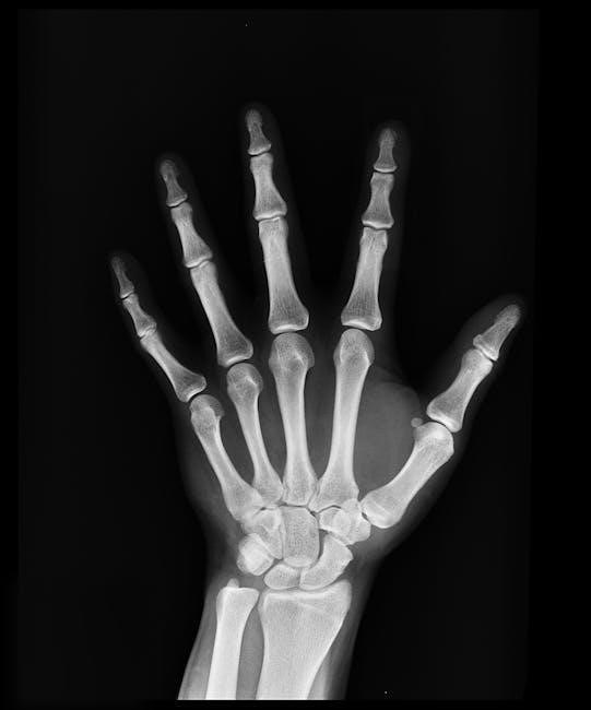
X-ray and fluoroscopy are foundational imaging modalities in radiology․ X-rays use ionizing radiation to produce static images of internal structures, while fluoroscopy provides real-time moving images․ Both are widely used for diagnosing bone fractures, lung diseases, and gastrointestinal issues․ X-rays are cost-effective and readily available, making them a first-line choice for many conditions․ Fluoroscopy, on the other hand, is particularly useful for guiding procedures and observing dynamic processes, such as swallowing or joint movements; These modalities are essential for quick and accurate diagnoses, offering valuable insights into patient conditions while being relatively non-invasive;
4․2 CT Scans
CT (Computed Tomography) scans are advanced imaging modalities that provide detailed cross-sectional images of the body․ They combine X-rays with computer technology to produce high-resolution visuals, enhancing diagnostic accuracy․ CT scans are particularly effective for evaluating trauma, detecting cancers, and assessing vascular diseases․ They are also used for guiding biopsies and drainage procedures․ Compared to traditional X-rays, CT scans offer superior detail, especially for soft tissues․ However, they involve higher radiation exposure, so their use is carefully considered․ Patient preparation may include fasting or contrast agents to enhance image clarity․ CT scans are versatile and play a critical role in emergency and complex diagnostic scenarios․
4․3 MRI (Magnetic Resonance Imaging)
MRI (Magnetic Resonance Imaging) uses magnetic fields and radio waves to produce detailed images of internal structures without ionizing radiation․ It excels in visualizing soft tissues, such as the brain, spinal cord, joints, and organs․ MRI is particularly useful for diagnosing neurological conditions, musculoskeletal injuries, and abdominal pathologies․ Patients with claustrophobia may require sedation․ Preparation involves removing metal objects and avoiding certain implants․ MRI is preferred for its high-resolution imaging and ability to detect early changes in tissues․ It is also valuable for assessing tumor extent and monitoring treatment response․ MRI is a critical tool in modern diagnostics, offering insights that other modalities cannot match․
4․4 Ultrasound
Ultrasound is a non-invasive imaging modality that uses high-frequency sound waves to produce real-time images of internal structures․ It is widely used for evaluating abdominal organs, pelvic structures, and musculoskeletal systems․ Ultrasound does not involve ionizing radiation, making it safe for pregnant women and children․ Common applications include fetal imaging, gallbladder assessments, and guiding biopsies or injections․ Doppler ultrasound also evaluates blood flow in vessels․ Patient preparation varies by exam, with fasting sometimes required for abdominal scans․ Ultrasound is portable, cost-effective, and provides dynamic imaging, enhancing diagnostic accuracy․ Its versatility and safety make it a preferred choice for numerous clinical scenarios, particularly in emergency settings and routine check-ups․
4․5 Nuclear Medicine
Nuclear Medicine uses small amounts of radioactive materials to diagnose and treat diseases․ It provides functional and metabolic information, often detecting issues before anatomical changes occur․ Common procedures include PET scans, bone scans, and thyroid studies․ PET scans combine with CT for detailed cancer staging․ Patient preparation varies, with some exams requiring fasting or specific timing․ Radiation exposure is minimal, and safety guidelines ensure low risk․ Nuclear Medicine is invaluable for assessing organ function, tumor activity, and infection sites․ It complements other imaging modalities, offering unique diagnostic insights that guide targeted therapies and improve patient outcomes across various clinical conditions and treatment monitoring․

Radiology Ordering Process
The Radiology Ordering Process involves selecting the appropriate imaging exam, preparing the patient, and ensuring accurate documentation․ It streamlines workflow, enhancing diagnostic efficiency and patient care quality․
5․1 Steps to Order a Radiology Exam
Ordering a radiology exam involves several key steps to ensure accuracy and efficiency․ First, assess the patient’s clinical needs to determine the appropriate imaging modality․ Next, review evidence-based guidelines to confirm the most suitable exam․ Then, prepare the patient by providing clear instructions on any necessary preparation, such as fasting or removing metal objects․ Enter the order through the healthcare system, including relevant clinical information․ Ensure proper documentation, including indications and patient history, to guide radiologists․ Finally, confirm the order and follow up to ensure the exam is completed and results are reviewed promptly․ Proper communication enhances diagnostic accuracy and patient care․
5․2 Choosing the Appropriate Imaging Modality
Selecting the right imaging modality is crucial for accurate diagnosis and patient care․ The Radiology Ordering Guide helps identify the most suitable exam based on clinical indications, patient symptoms, and imaging goals․ Factors such as the type of injury, disease, or condition guide the choice between modalities like X-ray, CT, MRI, or ultrasound․ For example, X-ray is ideal for bone fractures, while MRI is better for soft tissue injuries․ Patient-specific considerations, such as kidney function for contrast-enhanced CT scans, are also critical․ The guide ensures that the chosen modality balances diagnostic benefits with radiation exposure and patient safety, optimizing outcomes and reducing unnecessary procedures․
5․3 Role of Contrast Agents
Contrast agents are substances used in imaging to enhance the visibility of internal structures, improving diagnostic accuracy․ They are commonly administered via injection, ingestion, or other routes, depending on the modality․ For example, iodine-based agents are used in CT scans, while gadolinium is typical for MRI․ These agents highlight specific areas, such as blood vessels or tumors, aiding in precise diagnosis․ However, their use requires careful consideration of patient factors, including allergies, kidney function, and pregnancy status․ The Radiology Ordering Guide provides guidelines to ensure safe and effective use of contrast agents, balancing diagnostic benefits with potential risks to optimize patient outcomes․ Proper selection is critical to avoid complications and ensure clarity in imaging results․

Patient Considerations
Patient considerations in radiology focus on ensuring safety, comfort, and tailored care․ Factors like medical history, allergies, and physical condition guide decisions, optimizing outcomes while minimizing risks․
6․1 Patient Preparation for Imaging
Patient preparation for imaging ensures procedures are performed safely and effectively․ Clear instructions are provided, including fasting, medication management, and clothing requirements․ For MRI and CT scans, removing metal objects is crucial․ Ultrasound may require a full bladder․ Radiation exposure concerns are addressed, especially for pregnant patients․ Proper preparation reduces the need for repeat scans, improving efficiency and patient comfort․ RadiologyInfo․org offers detailed guides, helping patients understand their role in preparing for exams․ Open communication between patients and healthcare providers ensures all instructions are followed, optimizing diagnostic accuracy and minimizing risks․ Preparation varies by modality, emphasizing the importance of tailored guidelines for each imaging type․
6․2 Contraindications for Imaging
Contraindications for imaging are conditions or factors that make certain procedures unsafe or unsuitable for patients․ For example, MRI is contraindicated in patients with metal implants or pacemakers due to magnetic interactions․ Pregnant patients should avoid CT scans or X-rays to minimize radiation exposure risks․ Contrast agents used in CT or MRI may be contraindicated in patients with severe kidney disease or allergies․ Ultrasound is generally safer but may not be appropriate for certain conditions․ Understanding contraindications ensures patient safety and avoids complications․ Radiologists and referring physicians must assess each patient’s medical history to determine the most appropriate imaging modality․ This step is critical for minimizing risks and ensuring accurate diagnostics․
6․3 Special Populations (Pediatric, Geriatric, etc․)

Imaging considerations vary significantly for special populations, such as pediatric, geriatric, and pregnant patients․ Pediatric patients require lower radiation doses due to increased sensitivity, making ultrasound a preferred modality․ Geriatric patients may have mobility challenges or comorbidities, necessitating adapted imaging protocols․ Pregnant patients should avoid certain modalities like CT scans to prevent fetal harm․ Additionally, contrast agents may be contraindicated in patients with renal impairment․ Radiologists must tailor imaging strategies to these unique needs, ensuring safety and diagnostic accuracy․ Specialized guidelines and consultations are often essential to address these populations effectively, balancing clinical necessity with potential risks․

Legal and Ethical Aspects
The legal and ethical aspects of radiology ordering involve ensuring informed consent, radiation safety, and adherence to medico-legal standards․ Ethical responsibilities include minimizing radiation exposure and respecting patient autonomy while maintaining diagnostic accuracy and compliance with regulations․
7․1 Informed Consent for Radiology Procedures
Informed consent is a critical legal and ethical requirement in radiology, ensuring patients understand the nature, benefits, and risks of imaging procedures․ Patients must be fully aware of what the procedure entails, including potential side effects or complications, such as radiation exposure or allergic reactions to contrast agents․ Clinicians must also discuss alternative options and ensure patients have the capacity to make decisions․ The consent process must be documented, with patients signing a form after their questions are addressed․ This ethical practice respects patient autonomy and ensures transparency, aligning with legal standards and promoting trust in the healthcare provider-patient relationship․
7․2 Radiation Safety and Dose Optimization
Radiation safety and dose optimization are paramount in radiology to minimize patient exposure while maintaining diagnostic quality․ The “as low as reasonably achievable” (ALARA) principle guides efforts to reduce radiation doses without compromising image quality․ Techniques like adjusting X-ray beam settings, using shielding, and employing dose-reduction algorithms are essential․ Modern imaging systems incorporate features to optimize radiation exposure, particularly for vulnerable populations such as children․ Regular monitoring and staff training ensure adherence to safety protocols․ By prioritizing dose optimization, radiology professionals balance the benefits of imaging with the risks of radiation, ensuring safer care for patients․
7․3 Medico-Legal Considerations
Medico-legal considerations in radiology ordering emphasize the importance of proper documentation, informed consent, and adherence to privacy laws․ Accurate documentation of imaging orders and results is critical for legal protection and patient care continuity․ Informed consent ensures patients understand the risks and benefits of procedures․ Compliance with regulations like HIPAA protects patient confidentiality․ Clinicians must also be aware of liability risks associated with incorrect or delayed imaging orders․ Regular audits and adherence to ethical standards further mitigate legal challenges․ These measures ensure that radiology practices align with both medical and legal requirements, safeguarding patients and providers alike in the diagnostic process․

Technology in Radiology Ordering
Technology in radiology ordering involves Radiology Information Systems (RIS) and Picture Archiving and Communication Systems (PACS), streamlining exam orders and image management․ These tools enhance efficiency, reduce errors, and improve patient care by integrating with electronic health records (EHRs) and leveraging artificial intelligence for advanced image analysis and workflow optimization․
8․1 Radiology Information Systems (RIS)
A Radiology Information System (RIS) is a digital platform managing patient data, exam orders, and radiology workflows․ It integrates seamlessly with electronic health records (EHRs), enabling efficient scheduling and tracking of imaging procedures․ RIS streamlines communication between referring physicians and radiologists, ensuring accurate order entry and status updates․ Key features include patient registration, exam scheduling, and real-time tracking of procedure status․ RIS also supports billing processes and generates reports, enhancing operational efficiency․ By automating administrative tasks, RIS reduces errors and improves patient care, making it an essential tool in modern radiology departments․
8․2 Picture Archiving and Communication Systems (PACS)
Picture Archiving and Communication Systems (PACS) are digital platforms that store, retrieve, and display medical imaging data․ They integrate with Radiology Information Systems (RIS) and electronic health records (EHRs), enabling efficient access to patient images and reports․ PACS replaces traditional film-based systems, reducing storage costs and improving image accessibility․ It allows radiologists to view and analyze images from various modalities, such as X-rays, CT scans, and MRIs, in a standardized digital format․ PACS also supports image manipulation tools, enhancing diagnostic accuracy․ Secure access controls ensure patient data confidentiality․ By streamlining image management, PACS improves workflow efficiency and facilitates timely, accurate diagnoses in radiology departments․
8․3 Artificial Intelligence in Radiology
Artificial Intelligence (AI) is revolutionizing radiology by enhancing diagnostic accuracy and streamlining workflows․ AI-powered algorithms analyze medical images, detect abnormalities, and prioritize urgent cases, improving patient outcomes․ These systems leverage deep learning to identify patterns in imaging data, such as tumors or fractures, often with precision comparable to human radiologists․ AI also automates routine tasks, like image segmentation, freeing radiologists to focus on complex cases․ Integration with PACS and RIS systems enables seamless data flow, while ethical considerations, such as data privacy and transparency, remain critical․ By optimizing workflows and reducing diagnostic errors, AI is transforming radiology, making it faster, more efficient, and patient-centric․

Best Practices for Radiology Ordering
Adhering to evidence-based guidelines ensures appropriate imaging selection, minimizing unnecessary procedures․ Clear communication of clinical rationale and patient history enhances radiologist interpretation․ Regular updates and continuous learning are essential for optimal ordering practices․
9․1 Communication Between Referring Physicians and Radiologists
Effective communication between referring physicians and radiologists is crucial for accurate diagnosis and patient care․ Clearly conveying clinical context, patient history, and specific concerns ensures radiologists understand the rationale behind imaging requests․ Using precise terminology and avoiding vague language helps radiologists prioritize and interpret images effectively․ Regular updates and feedback loops enhance collaboration, ensuring timely and relevant reporting․ Leveraging tools like Radiology Information Systems (RIS) and Picture Archiving and Communication Systems (PACS) streamlines this process, reducing errors and improving efficiency․ Open dialogue fosters trust and ensures that imaging aligns with clinical needs, ultimately benefiting patient outcomes․
9․2 Documentation and Reporting
Accurate and detailed documentation is essential for effective radiology reporting․ Radiologists must document all relevant findings, ensuring clarity and conciseness․ Reports should include patient history, clinical context, and prior imaging comparisons to aid referring physicians․ Standardized reporting formats improve consistency and readability, highlighting abnormal findings and recommended next steps․ Timely communication of critical results is vital to prevent delays in patient care․ Additionally, documentation should comply with legal and ethical standards, maintaining patient confidentiality and professionalism․ Clear and precise reporting fosters better collaboration between radiologists and referring physicians, ultimately enhancing diagnostic accuracy and patient outcomes․ Proper documentation also supports quality improvement initiatives and legal compliance․
9․4 Continuous Learning and Updates
Continuous learning and updates are crucial in radiology to stay current with advancements in imaging technologies and evidence-based guidelines․ Radiologists and referring physicians must engage in ongoing education to ensure they are aware of the latest modalities, techniques, and clinical recommendations․ Regular updates to the Radiology Ordering Guide reflect new research, improving the accuracy of imaging selections․ Leveraging resources like webinars, journals, and professional workshops helps maintain proficiency․ Additionally, incorporating feedback from patient outcomes and technological advancements ensures the guide remains relevant and effective․ Continuous learning fosters a culture of improvement, enhancing diagnostic accuracy and patient care while adapting to evolving medical standards․
The Radiology Ordering Guide is an essential tool for improving imaging decisions, ensuring patient safety, and optimizing diagnostic outcomes․ Its continuous evolution aligns with advancing technologies and evidence-based practices․
10․1 Summary of Key Points
The Radiology Ordering Guide serves as a comprehensive resource for standardizing imaging decisions, ensuring they align with clinical needs and patient safety․ By emphasizing evidence-based recommendations, it helps providers choose the most appropriate imaging modalities, reducing unnecessary exams․ The guide also highlights the importance of collaboration between referring physicians and radiologists, fostering clear communication and accurate diagnoses․ Its focus on patient-centered care ensures considerations for special populations and safety measures like radiation dose optimization․ Ultimately, the guide enhances resource utilization, improves outcomes, and adapts to advancing technologies, making it an indispensable tool for modern healthcare practices․
10․2 Future Directions in Radiology Ordering
The future of radiology ordering lies in advancing technologies like AI and machine learning, which will enhance decision-support systems, enabling more precise and personalized imaging recommendations․ Integration with electronic health records (EHRs) will streamline workflows, improving efficiency and reducing errors․ The use of predictive analytics will help anticipate patient needs, optimizing care pathways․ Additionally, advancements in imaging modalities and contrast agents will expand diagnostic capabilities․ Patient-centered approaches will prioritize safety, minimizing radiation exposure while ensuring high-quality outcomes․ These innovations will transform radiology ordering, making it more accessible, efficient, and aligned with evolving clinical demands, ultimately improving patient care and operational excellence in healthcare settings․